
General Eye Care
Desert Vision Center does not accept new patients for routine eye care. Dr. Tokuhara specializes in complex diseases of the eye and does not perform routine eye screening examinations for healthy eyes. Unlike other offices, Dr. Tokuhara has an open door policy for any of his surgery patients. When you chose Dr. Tokuhara for your cataract surgery, you have an eye doctor for life. At Desert Vision Center, we provide general eye care for our surgery patients after their surgery or patients who are developing a vision threatening eye problem. We do not provide post-operative nor general eye care for patients whom have had eye surgery elsewhere.
The Normal Eye
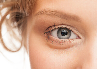
The human eye is truly amazing. It focuses light to form images or “pictures” at the back of your eye, much like a camera. The eye instantly changes these images into electrical signals and sends them to your brain. The brain interprets the signals and you experience “seeing”. The eye accommodates to changing lighting conditions and focuses rays of light originating from various distances.
What Are the Structures of the Eye?
Cornea: This clear outer lens provides two-thirds of the focusing power of the eye. The cornea is made up of transparent tissue, which allows light to pass through. The cornea focuses the light by bending it so the light rays form an image on the retina. Since the cornea has the greatest bending (focusing) power, it is the cornea’s shape that determines a great deal of quality of your vision.
Iris & Pupil: The colored part of the eye is called the iris and functions much like the iris of a camera, opening, and closing, to control the amount of light entering through the pupil (that dark opening in the center the iris).
Crystalline lens: The crystalline lens is located behind the iris and provides one third of the focusing power of the eye. The crystalline lens works to further bend light rays as they pass through the eye to form an image on the retina.
Retina: Located in the lining at the back of the eye, the retina acts as an electrical system to send impulses to your brain via the optic nerve. The retina contains photoreceptor cells that collect information from light as it passes through the cornea and crystalline lens to the back of the eye. Your brain interprets the retina’s electrical response into what you to experience as images.
Fovea: The focal point at the center of the retina is called the fovea. Light-focused here produces the sharpest vision.
Vision
The eye focuses by bending incoming light rays to meet at a single point. Ideally, this single point lands directly on the fovea, the central point of the retina. If the light rays reach this perfect placement, you experience clear, sharp images.
However, if the focal point is behind the retina or in front of the retina, the image on the retina will not be fully formed and will be interpreted by your brain as blurred. This is very much like focusing a projector onto a blank movie screen. If the projection is too close or too far from the screen, the images are blurred. Set at the correct distance, you may enjoy the show!
Dr. Tokuhara will determine whether you are nearsighted, farsighted, astigmatic, and/or presbyopic by measuring where your eye focuses light. Depending on your refraction and anatomy, he will discuss different treatment options with you.
Nearsightedness (myopia)
Nearsighted individuals typically have problems seeing well at a distance and are forced to wear glasses or contact lenses. The nearsighted eye is usually longer than a normal eye, and its cornea may also be steeper. Therefore, when light passes through the cornea and lens, it is focused in front of the retina. This will make distant images appear blurred.
Farsightedness (hyperopia)
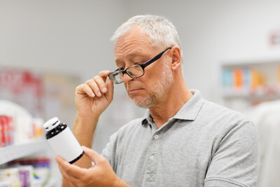
Farsighted individuals typically develop problems reading up close before the age of 40. The farsighted eye is usually slightly shorter than a normal eye and may have a flatter cornea. Thus, the light of distant objects focuses behind the retina unless the natural lens can compensate fully. Near objects require even greater focusing power to be seen clearly and therefore, blur more easily.
Astigmatism
Asymmetric steepening of the cornea or natural lens causes light to be focused unevenly, which is the main optical problem in astigmatism. To individuals with uncorrected astigmatism, images may look blurry or shadowed. Astigmatism can accompany any form of refractive error and is very common. Astigmatism can be corrected with glasses, contact lenses, corneal relaxing incisions, and special implant lenses.
Presbyopia
Presbyopia is a condition that typically becomes noticeable for most people around age 45. In children and young adults, the lens inside the eye can easily focus on distant and near objects. With age, the lens loses its ability to focus adequately.
Although presbyopia is not completely understood, it is thought that the lens and its supporting structures lose the ability to make the lens longer during close vision effort. To compensate, affected individuals usually find that holding reading material further away makes the image clearer. Ultimately, aids such as reading glasses are typically needed by the mid-forties. Besides glasses, presbyopia can be dealt with in a number of ways. Options include: monovision and multifocal contact lenses, and new presbyopia correcting implant lenses.
Color Blindness
The most common form of color blindness is red-green color deficiency. 8% of men and 0.6% of women have the red-green form of color blindness because of X-linked genetic inheritance. Colored tablets or diagrams (such as the Ishihara Test) are used to test for color vision blindness.
At Desert Vision Center, we offer new solutions for color blindness with Enchroma Glasses. The glasses help people with color vision see the beautiful world the way it is meant to be seen.
We’ve had fantastic results and stories from patients trying on the new glasses for the first time in our office. Do you have color blindness? Take the two minute online color vision test. and find out here.
Eye Examination
An eye examination includes a review of your medical history, an analysis of your visual acuity, which may or may not include a prescription for glasses, an examination of the eye’s physical well-being, content and surrounding tissue, and an evaluation of the eye’s connection with the visual system inside your brain.
Who needs an eye examination?
An adult examination may require eyeglass measurement (refraction) for optimal visual acuity, screening for glaucoma, analysis of eye structures and surrounding tissues, and recommendations for treatment as necessary. Adult examinations look for diseases that can be treated or prevented so that sight can be restored or protected from further or future loss of vision.
How often should I have my eyes checked?
Adults with normal findings on an initial examination should have their eyes checked every two to four years, depending on age.
| Age | Eye Exam |
| 20-39 | Every 3 – 5 years |
| 40-64 | Every 2 – 4 years |
| 65 or older | Every 1 – 2 years |
Adults who are being treated for specific eye conditions will need to return for follow up more frequently and we will provide you with a specific time interval when you should return. We will schedule this appointment for you if you wish, or can provide you with a reminder card that will be mailed to you prior to when you should be seen again.
How is an eye examination performed?
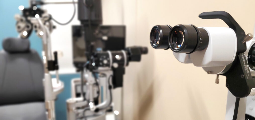
Dr. Tokuhara uses the most modern testing equipment that allows us to carefully screen for refraction. Before an eyeglass prescription is written, this screening information is retested by one of the doctors to make sure it is correct. Glaucoma testing includes a measurement of eye pressure. Ocular alignment is evaluated for cross-eyed or other eyestrain caused by strabismus (eyes are not aligned and there is a stress in the ability of the two eyes to view the same target). A careful analysis of the external structures around the eyes is performed, including the eyelid, lashes, tear drainage, and eye moisture. A careful analysis of the internal structures of the eye includes the cornea, iris, pupil, lens, vitreous, retina, optic nerve, and blood vessels.
As necessary, further testing is performed, which may include measurement of actual tear production, cell thickness and condition of the cornea, an analysis of risk of glaucoma (gonioscopy), a detailed evaluation of peripheral vision or visual field, a quantitative and qualitative measurements of the optic disc and retina, and wavefront analysis of internal and external ocular tissue.
Is a refraction (measurement for eyeglass prescription) a part of every examination?
Our job is to evaluate and care for people’s eyes. As part of this, we must know how well you see. To accomplish this, we must do enough of a refraction to score a person’s visual acuity. Knowing how well a person sees and obtaining a basic knowledge of a person’s general refraction is done on nearly every patient who comes to our office except in an emergency, or a few other situations when we may not be initially able to obtain this information.
What will happen to my eyes during my examination?
Most adult patients will be given eye drops that allow us to check for glaucoma. These anesthetic eye drops sting a little for a few moments and then quickly wear off.
Patients will be given “dilating drops” that open the pupils widely so that we can evaluate the back of the eye and so that we can measure the eyeglass refraction most precisely. These dilating drops may cause some loss of near vision/reading vision for a few hours. Dilating drops may also cause significant difficulty in bright sunlight.
Why do you have to do all of this?
In order to take care of you to the best of our ability, and in order to be as thorough as possible in examining you, all of the tests and eye drops are necessary. Should you have any question about this, please ask one of our fine ophthalmic technologists or Dr. Tokuhara. Your sight is precious; we want to do the best job possible taking care of you.
DVC Promise
Dr. Tokuhara performs advanced technology diagnostic testing and treatment, as well as taking the time necessary to provide each patient with information needed to fully understand his or her condition and to achieve the best possible visual outcome.